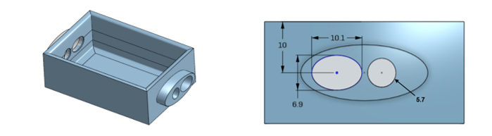
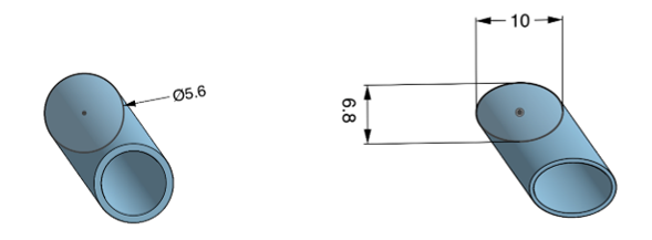
To simulate the cannulation access points, 3D moulds have been designed resembling each point. After designing and printing, each will be filled with a substance called EcoFLEX, which will harden after a period of time and creating pads that represent the cannulation access points. EcoFLEX is a flesh-like material which aids the realism constraint in the design, more importantly when using EcoFLEX ultrasound scan is possible. The ability to have ultrasound scanning is trivial when performing cannulation, as well as for checking the blood flow. The design of the 3D moulds for the jugular cannulation site represented in the below figures with the overall structure and detailed dimensions.
 The design of the 3D moulds for the heart cavity pad are represented in the below figures.
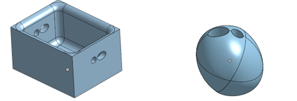
The 3D moulds have been designed with specific dimensions according to the information observed from Table 2-3. To simulate the access point for the jugular cannulation site, the two simulated vessels will be inserted inside the designed mould represented in Figure 3-3. Thus, EcoFLEX material will fill the final mould to represent a realistic jugular cannulation site. However, simulating the heart cavity will be slightly different. First, EcoFLEX material will fill the heart container (represented in Figure 3-4 (a)) up to 10mm and wait until it hardens. Afterwards, the simulated heart will be inserted inside the container along with the two vessels inside both, the heart and the container. Then, EcoFLEX material will fill the final mould and wait until it hardens. After that, EcoFLEX material will be extracted from the mould. Next, the pad will be cut in half to extract the simulated heart form the material. Finally, the pad will be reattached together by a very thin layer of EcoFLEX, to represent a realistic neonate heart cavity model.
However, one of the main feedback points that the doctor stressed on after the demo that was held during the first HMC visit, was that the pads used to simulate the cannulation sites had air bubbles inside of them, which disturbed the ultrasound scan and affected their clarity. So, the use of a different ultrasoundable material was proposed. One material that could be a possible alternative is “agar phantoms”. Agar phantoms is an ultrasoundable material categorized under the tissue mimicking materials (TMM), where they are commonly used to mimic or represent soft tissue such as the blood vessels that have to be simulated in this design [16].
EcoFLEX material is set to be used to represent the cannulation sites for the proposed design. However, if the air bubbles dilemma is not fixed, the help of chemical engineers will be required to prepare agar phantoms and use it to mimic the cannulation sites in this design. Substituting the EcoFLEX with agar phantoms in the design to represent the cannulation access points will not affect the designing process of the moulds. Since, both martials could mimic the shape of the mould the same way.