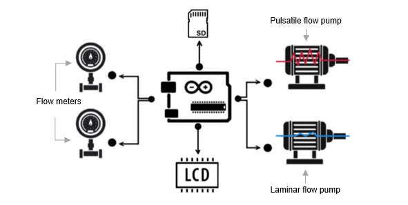
A block diagram of the embedded system is presented in the below figure. The embedded system consists of several components, each component has a different functionality aiding the designed system requirements. The microcontroller has the significant part in the design, it is the “brain” of the system that controls the programmable pumps for the blood circulation, the flow meters for measuring the blood flow rate and to display on the LCD screen and the normal or the abnormal EEG signals prestored on the SD card, to achieve the objective of adding emergency situations on the simulator. Arduino MEGA is the microcontroller that has been chosen for the proposed design. It has been chosen because of its large memory capacity; where it has 256 KB of flash memory for storing codes [20]. Moreover, it has 54 digital pins that can be used as an input or output, it has also 16 analog inputs that can provide 10 bits of resolution for each pin [20]. All these pins can be connected to the system components to perform the process of the system.

After implementing the system, the SD card and the LCD screen will be connected to the microcontroller through a SD card module and an LCD screen shield. Actually, the LCD screen has exactly the same size of the microcontroller with the same number of pins, where it can be directly attached to the microcontroller. However, it should be connected to the LCD screen shield first, to give the microcontroller pins some space for other components to be connected as well. The below figure shows the schematic diagram of the SD card and the LCD screen connections.

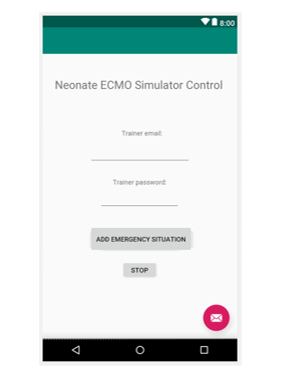
Moreover, a dataset of normal and abnormal EEG signals will be stored on the SD card. Thus, signals will be displayed on the LCD screen monitor for the trainee to observe during the cannulation procedure. This feature will be controlled by the trainer through a mobile application, to trigger a seizure. Therefore, a simple mobile application layout has been designed by using Android Studio Software, and the final design of the mobile application will be integrated into the main mobile application that has been developed earlier by the first team that designed the ECMO machine simulator. Figure below represent the GUI of the desired mobile application.

The trainee should always check the displayed signals for any abnormal brain activity. Integrating EEG signals into the system gives it credibility, where it will reflect more complex realistic situations for the trainees to act on, and thereby be prepared for real-life situations.
A block diagram represented in the below figure, for the neonatal ECMO cannulation simulator, to clarify the process of the system.
